
Little Brains, Big Discoveries: How Brain Organoids Are Transforming Neuroscience
By: Likhitha Selvan
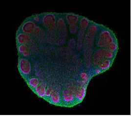
Cross-section of a two-month-old cerebral organoid observed under a fluorescence microscope
Imagine trying to understand how a city develops without ever seeing its construction. For decades, this was the challenge faced by neuroscientists studying the human brain. Researchers could examine adult brain tissue or rely on animal models, but observing the earliest stages of human brain development was nearly impossible. In the last decade, however, scientists have developed a novel solution by the name of brain organoids. Brain organoids are small three-dimensional clusters of neural tissue grown from human stem cells that mimic aspects of the developing brain. These miniature models are transforming neuroscience by allowing researchers to investigate brain development, neurological disease, and drug responses in ways that were previously impossible.¹
Brain organoids are created using human pluripotent stem cells, cells thathave the ability to differentiate into nearly any cell type in the body. When scientists expose these cells to specific chemical signals and grow them in a three-dimensional matrix, they begin differentiating into neurons and supporting glial cells, a group of non-neuronal cells that support and protect neurons in the brain. Over time, these cells self-organize into layered structures resembling the early cerebral cortex, the outer region of the brain responsible for functions such as perception, memory, decision-making, and voluntary movement.¹
This self-organization is one of the most fascinating aspects of organoid biology. Instead of scientists building brain tissue piece by piece, the cells themselves follow developmental programs encoded in their DNA. Early breakthroughs in this field came from researchers who demonstrated that stem cells could form cerebral organoids capable of modeling early human brain development. This included the formation of neural progenitor zones, regions that contain rapidly dividing precursor cells that generate neurons in addition to primitive cortical layers, the early stacked layers of neurons that later mature into the organized structure of the cerebral cortex.² These models allowed scientists to observe developmental processes that occur during the first weeks of brain formation.
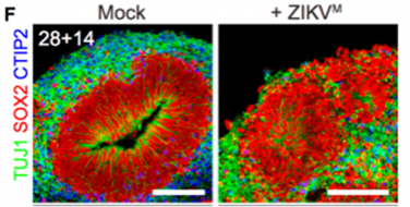
Experimental infection of human brain organoids with Zika virus demonstrating reduced neural progenitor cell survival and impaired growth.
One innovative use of brain organoids emerged during the Zika virus outbreak in between 2015 and 2016. Doctors noticed that babies born to infected mothers frequently developed microcephaly, a condition characterized by an abnormally small brain. Researchers used human brain organoids to investigate how the virus affected neural development. When the organoids were exposed to Zika virus in laboratory experiments, scientists observed that the virus specifically infected neural progenitor cells, the cells responsible for producing neurons in the developing brain. This infection caused extensive cell death and dramatically reduced organoid growth, providing direct evidence that the virus disrupts brain development by targeting these progenitor cells.³
These experiments demonstrated how organoids could be used to model diseases that are otherwise difficult to study. Unlike animal models, which often differ significantly from human biology, organoids are derived from human cells and therefore capture species-specific features of development. Researchers have since used brain organoids to study a wide range of neurological conditions, including Alzheimer’s disease, Parkinson’s disease, and autism spectrum disorders.⁴
For example, scientists have developed midbrain organoids to study Parkinson’s disease. These organoids contain dopaminergic neurons, the type of neuron that degenerates in Parkinson’s patients. Researchers have observed that these neurons accumulate pathological α-synuclein proteins and show signs of cellular stress, mimicking the early stages of the disease.⁴ These findings have added to scientists’ current knowledge on how neurodegeneration begins and progresses.
Brain organoids have also become essential tools for studying genetic neurodevelopmental disorders. In one such study, researchers used organoids derived from patients with Rett syndrome, a neurological disorder caused by mutations in the MECP2 gene. Scientists observed abnormal neuronal signaling and reduced synaptic connectivity in these organoids. When experimental drugs were applied, some of these cellular abnormalities were partially reversed, suggesting potential therapeutic strategies.⁵
Beyond modeling disease, scientists are beginning to construct more complex systems by combining multiple organoids together. These structures, known as assembloids, allow researchers to study how different brain regions communicate with one another. By fusing organoids representing different regions of the brain, scientists can observe processes such as neuron migration and circuit formation, which is essential to neurodevelopmental disorders.¹
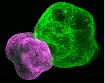
Cortico-striatal assembloid formed by the fusion of two brain organoids.
Despite these advances, organoids are still far from replicating the full complexity of the human brain. They lack blood vessels, immune cells, and the intricate architecture of a mature brain. As a result, they remain simplified models of early brain development rather than miniature functioning brains. Even so, researchers continue to refine organoid systems by introducing vascular cells, improving nutrient delivery, and extending the time organoids can survive in culture.
As these models grow more complex, they also raise ethical questions. Some scientists have wondered whether sufficiently advanced organoids might eventually develop complex neural activity resembling aspects of what might be considered consciousness. Current evidence suggests that existing organoids remain far too simple to support such processes, but the discussion emphasizes the importance of ethical oversight as the technology evolves.⁴
Brain organoids represent one of the most exciting innovations in modern neuroscience. By recreating key stages of human brain development in the laboratory, these tiny clusters of cells allow researchers to investigate diseases, test potential therapies, and uncover the biological mechanisms that shape the brain. From explaining how viruses like Zika damage developing neurons to modeling disorders such as Parkinson’s and Rett syndrome, organoids have already transformed how scientists study the human nervous system.
Though only a few millimeters in size, these miniature models offer a magnified look into the earliest stages of human brain development. As organoid technology continues to advance, it may help scientists unlock new treatments for neurological disease and deepen our understanding of the most complex organ in the human body.
References
1. Sun, Y.; Paşca, S. P.; Sloan, S. A. Brain Organoids: A New Paradigm for Studying Human Brain Disorders. Front. Neurosci. 2025, 19, 1699814.
2. Lancaster, M. A.; Knoblich, J. A. Generation of Cerebral Organoids from Human Pluripotent Stem Cells. Nature 2014, 501, 373–379.
3. Garcez, P. P.; Loiola, E. C.; Madeiro da Costa, R.; et al. Zika Virus Impairs Growth in Human Brain Organoids. Science 2016, 352, 816–818.
4. Shima, S.; et al. Human iPSC-Derived Neural Organoids for Modeling Neurodegenerative Diseases. Trends Neurosci. 2025, 48, 152–165.
5. Mariani, J.; Coppola, G.; Zhang, P.; et al. FOXG1-Dependent Dysregulation of GABAergic Neurons in Autism Spectrum Disorders. Cell 2015, 162, 375–390.